Info
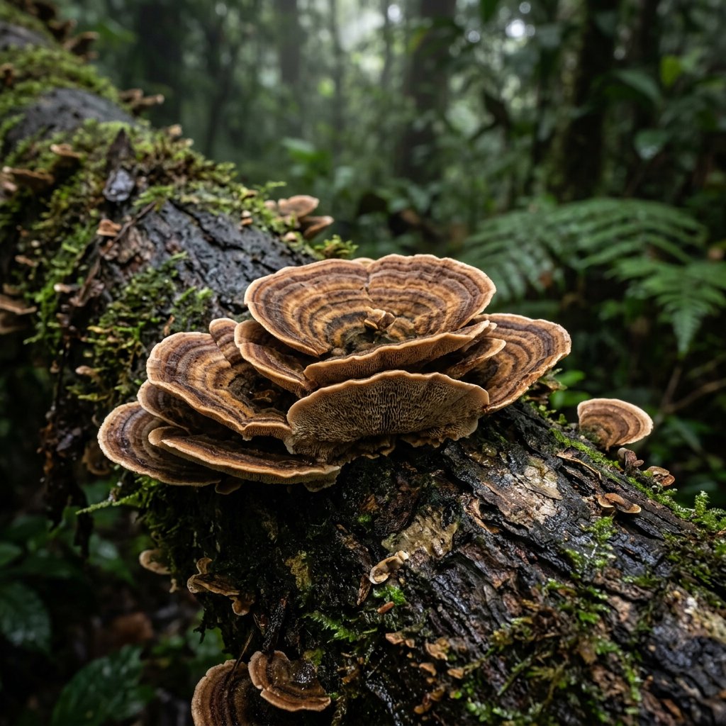
Often found clinging to fallen branches like tiny, colorful ears, Microporus affinis is a stunning example of a "bracket fungus." Known for its vibrant, banded patterns, it looks like a miniature piece of forest art decorating the undergrowth. While it lacks a single common name, it is frequently admired by hikers in tropical regions for its elegant, fan-like symmetry and velvet-soft texture.
🔍 How to Identify
- 🍄 The Cap: A thin, leathery, fan-shaped bracket featuring striking concentric rings of color. These bands range from deep chocolate brown and ochre to creamy yellow and stark white.
- 🕳️ The Pores: The underside is remarkably smooth to the naked eye. This is because the pores are microscopic (hence the name Microporus), appearing as a pale, cream-colored surface.
- 🪵 The Stem: Unlike many shelf fungi that grow flush against wood, this species usually has a short, stiff, lateral stem (stipe) that attaches it to its host branch.
🌲 Habitat & Ecology
- 🌳 The Forest Recycler: This fungus is a saprobe, meaning it feeds on dead organic matter. It is a "white rot" fungus, specialized in breaking down the tough lignin in hardwood, which helps recycle nutrients back into the forest floor.
- 🌏 Tropical Preference: It is most commonly found in the humid tropical and subtropical forests of Asia, Africa, and the Pacific. It thrives on fallen logs and decaying branches where moisture is consistent.
⚠️ Safety & Toxicity
- 🚫 WARNING: While Microporus affinis is not known to be a deadly "poisonous" mushroom, it is strictly inedible.
- 🦷 Tough Texture: The flesh is extremely leathery and cork-like. It does not soften with cooking and would be impossible to chew or digest, offering zero culinary value.
- 🤚 General Advice: It is safe to touch for identification purposes, but it is a best practice to wash your hands after handling any wild fungi to avoid any potential irritation or accidental ingestion of spores.
✨ Fun Fact
🔍 Hidden Details: The pores on the underside are so tiny that there are typically 8 to 10 of them packed into a single millimeter! To see the architecture of where the spores are released, you almost always need a high-powered magnifying glass or a microscope.
More Details